Flow Cytometry
October 11, 2011
Just as
French was the "
lingua franca" of
law and
diplomacy in past years,
Latin and
Greek are used by
physicians and
medical researchers so they can be understood internationally. Few people today are
classical scholars, but many people recognize quite a few of the Greek words in the medical
lexicon.
When confronted by any word containing "cyt," we know that it concerns
cells. It derives from the Greek word, κυτος, transliterated as kytos, that means "a hollow." The hollow aspect was readily apparent to
Robert Hooke when he observed the structure of
cork under a
microscope (see figure). The shapes reminded him of the small rooms, called cells, where
monks lived.
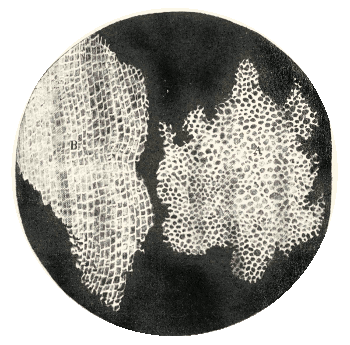
Drawing of cork cells, as viewed by Robert Hooke under a microscope.
"...Our microscope informs us that the substance of Cork is altogether fill'd with Air, and that the Air is perfectly enclosed in little Boxes or Cells..."[1]
From Hooke's Micrographia, via Wikimedia Commons)
The number of
blood cells of various types could only be counted by use of a microscope prior to the invention of the modern method of cellular analysis by the
electrical engineer,
Wallace Coulter.[2] Coulter's eponymous
Coulter counter was a method of electrically classifying particles suspended in a
conductive solution as they flowed through a thin tube from one chamber to another (see figure).
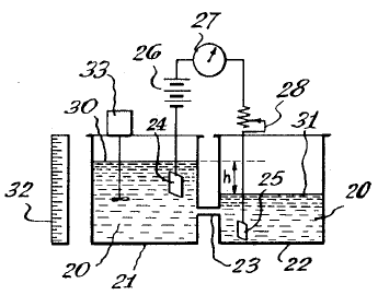
Figure one of US Patent No. 2,656,508, "Means For Counting Particles Suspended In A Fluid," by Wallace H. Coulter, October 20, 1953.
(via Google Patents))
Using blood cells as an example, a dilute mixture of blood in an
isotonic solution flows through a thin tube from one reservoir to another. The isotonic solution is
electrically conductive, so a current will flow between an electrode in one reservoir to an electrode in the other.
When a blood cell enters the tube, it blocks a fraction of the
current flow in the tube. Since the reduction in current depends on the
cross sectional area of the cell, the cell size can be inferred, and different cell types can be counted. Thus, a
red blood cell having a diameter of about 6-8
μm and thickness of 2 μm can be distinguished from the smaller
platelets (about 2-3 μm diameter) and the larger
white blood cells (7-21 μm diameter).
Although Coulter's original detection scheme used electrical current for detection, modern
flow cytometers have many modern detection technologies available.
Optical techniques are the most common, and this leads to sensitive analyses when
fluorescent tags can be attached to cells.
It's even possible to sort cells.
Charged droplets containing individual cells are steered one way, or another, by
electric fields that are controlled by the sensed cellular size.[3] In a
previous article, I wrote about a variation of the Coulter method for
sequencing DNA.[4]
Nowadays, the
electronic circuitry for flow cytometry so easy to implement that any engineer experienced in
embedded systems design can roll his own quite cheaply. Things are easier still if you can append your device to a common electronic device. That's what researchers at
UCLA's Henry Samueli School of Engineering and Applied Science have done in converting a
cellphone to a flow cytometer at minimal expense.[4-5]
The team, led by
Aydogan Ozcan, a professor of electrical engineering and bioengineering at UCLA, fabricated this eighteen
gram device from a handful of inexpensive parts that included a simple
lens ($3), a plastic color filter ($1), two
light-emitting diodes ($0.30/each), some
optofluidic piping, and
batteries.[5] The optofluidic components are a
syringe pump and a disposable
microfluidic channel through which the cells flow. The device is for use with fluorescently tagged cells, and it uses the
cellphone camera as the optical detector.[4-5]
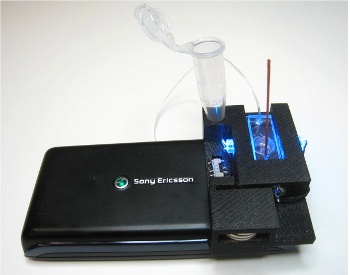
Cytometry attachment for a cellphone. The cellphone camera is used to image the flowing cells for analysis.
(UCLA Image/Aydogan Ozcan).[5])
The cellphone camera records video of the cells as they stream by, and
computer analysis counts the labeled cells. The spatial resolution is two μm, and the system was tested by measuring the volumetric density of human white blood cells (7-21 μm diameter).[4-5] Says Ozcan,
"We have more than 5 billion cell phone subscribers around the world today, and because of this, cell phones can now play a central role in telemedicine applications... Our research group has already created a very nice set of tools, including cell phone microscopes, that can potentially replace most of the advanced instruments used currently in laboratories."[5]
The
group's web site shows other innovative analysis equipment.[6] The cellphone cytometer is intended for such uses as monitoring the health of
HIV patients and examining
water-quality in poorer countries.[4-5] The cellphone cytometer study was funded by the
National Institutes of Health, the
National Science Foundation, the
Office of Naval Research, the
Bill & Melinda Gates Foundation and the the
Vodafone Americas Foundation.[5]
References:
- Robert Hooke, "Micrographia: or, Some physiological descriptions of minute bodies made by magnifying glasses," (J. Martyn & J. Allestry, London, 1665), p. 112f.
- Wallace H. Coulter, "Means For Counting Particles Suspended In A Fluid," US patent No. 2,656,508, October 20, 1953.
- M. J. Fulwyler, "Electronic Separation of Biological Cells by Volume," Science, vol. 150, no. 3698 (November 12, 1965), pp. 910-911.
This Blog, "DNA Analysis," September 17, 2010.
- Hongying Zhu, Sam Mavandadi, Ahmet F. Coskun, Oguzhan Yaglidere and Aydogan Ozcan, "Optofluidic Fluorescent Imaging Cytometry on a Cell Phone," Analytical Chemistry, vol. 83, no.17 (September 1, 2011), pp 6641-6647.
- Wileen Wong Kromhout, "Got flow cytometry? All you need is five bucks and a cell phone," UCLA Press Release, July 26, 2011.
- Ozcan Research Group Web Site.
Permanent Link to this article
Linked Keywords: French; lingua franca; International law; diplomacy; Latin; Greek; physician; medical research; classical scholar; lexicon; cell; Robert Hooke; cork; microscope; monk; Micrographia; Wikimedia Commons; blood cell; electrical engineer; Wallace Coulter; Coulter counter; electrical conductor; conductive; solution; Google Patents; isotonic solution; current flow; cross sectional area; red blood cell; μm; platelet; white blood cell; flow cytometer; optical; fluorescent tag; charge; electric field; DNA sequencing; electronic circuitry; embedded system; UCLA's Henry Samueli School of Engineering and Applied Science; cellphone; Aydogan Ozcan; gram; lens; light-emitting diode; optofluidic; battery; syringe pump; microfluidic channel; cellphone camera; computer analysis; HIV; fecal coliform bacteria; water-quality; National Institutes of Health; National Science Foundation; Office of Naval Research; Bill & Melinda Gates Foundation; Vodafone Americas Foundation; US patent No. 2,656,508.